








US Based · Spatial Transcriptomics Services
Spatial Transcriptomics Services
Map cellular function directly within tissue architecture with FFPE or fresh-frozen tissue from a suite of certified high-resolution spatial transcriptomics and in situ profiling services in as little as 2 to 4 weeks.
Spatial services
Multi-Platform Workflows for Tissue Architecture
10x Genomics Visium
NGS-based spatial transcriptomics
Unbiased whole-transcriptome tissue mapping with integrated H&E morphology and Illumina sequencing data.
10x Genomics Xenium
Subcellular in situ profiling
High-throughput in situ spatial analysis with subcellular resolution for targeted gene expression and protein analysis.
Illumina StrataMap
Cellular transcriptome discovery
Our newest spatial assay delivers unbiased whole-transcriptome capture with the largest capture area for deeper gene discovery.
Not sure which spatial platform fits your project? Our scientists provide complimentary technical consultations to help you select the optimal workflow based on your sample quality, block availability, and research objectives. Read more about our services below or use the shortcut to explore the Technical Documents and FAQ sections.
The Signios Advantage
Workflow Performance & Technical Excellence
From block sectioning to final spatial alignment, our optimized tissue-handling protocols maximize transcript yield, protect RNA integrity, and preserve clean morphology for every project.
![]()
End-to-End Solutions
Integrated workflows spanning histological block preparation, TMA building, H&E imaging, spatial mapping, NGS sequencing, and comprehensive data reporting in SF Bay Area lab.
![]()
Multi-Platform Profiling
Access a comprehensive spatial biology portfolio featuring whole-transcriptome capture, high-plex targeted in situ imaging, and high-density resolution to your study boundaries.
![]()
Unrivaled Sensitivity
Validated tissue transfer and capture protocols maximize transcript yield, protecting low-input or thin cryosections as well as highly challenging, degraded clinical archived specimens.
![]()
High Structural Preservations
Certified histology pipelines utilize precision cryosectioning and microtomes to prevent tissue detachment or degradation, delivering crisp, publication-grade morphology overlays.
![]()
US-Based Services
All tissue sectioning, slide placement, high-resolution imaging, and library sequencing are performed in-house at our Foster City, CA facility. No outsourcing or international shipping.
![]()
Expert Support
Every spatial biology project is overseen end-to-end by PhD-level scientists providing dedicated guidance on tissue quality assessments (DV200 verification), custom gene panel design and platform guidance.
![]()
Scalable Infrastructure
Powered by the latest Illumina NovaSeq X+ sequencing alongside advanced spatial analyzers for ultra-deep molecular depth and high spatial resolution throughput.
![]()
Fast Turnaround Times
Optimized laboratory protocols utilizing state-of-the-art automation and processing systems to ensure efficient slide handling and rapid data delivery in 1-4 weeks.


The Signios Partnership
Laboratory Workflow & Quality Assurance
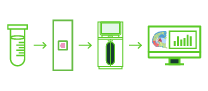
A transparent, five-step workflow designed to support TMA block building, QC checks and slide placement to preserve tissue morphology and ensure exact spatial data alignment.
Step 1
Our scientists provide guidance to align our spatial technical workflows (Visium, Xenium, or StrataMap) with your unique sample dimensions, tissue preparation states, and research objectives.
Step 2
Register your FFPE blocks, fresh-frozen tissues, or pre-cut sections via our streamlined customer portal. Schedule a local Bay Area pick-up or ship directly via verified cold-chain logistics.
Step 3
Our histology team performs rigorous tissue quality control, providing formal morphology, RNA Integrity Number (RIN), or DV200 reports and confirmation of slide placements before processing.
Step 4
Slides undergo precision mounting, automated probe hybridization or matrix capture, and standard high-resolution histology imaging, transitioning seamlessly to our advanced high-throughput imaging and sequencing instruments.
Step 5
Secure delivery of analysis-ready spatial files and QC reports via secure transfer. Advanced automated pipelines (including Space Ranger, Xenium Ranger, and DRAGEN Spatial) are utilized for interactive downstream spatial differential expression interpretation.
Our scientists provide guidance to align our spatial technical workflows (Visium, Xenium, or StrataMap) with your unique sample dimensions, tissue preparation states, and research objectives.
Register your FFPE blocks, fresh-frozen tissues, or pre-cut sections via our streamlined customer portal. Schedule a local Bay Area pick-up or ship directly via verified cold-chain logistics.
Our histology team performs rigorous tissue quality control, providing formal morphology, RNA Integrity Number (RIN), or DV200 reports and confirmation of slide placements before processing.
Slides undergo precision mounting, automated probe hybridization or matrix capture, and standard high-resolution histology imaging, transitioning seamlessly to our advanced high-throughput imaging and sequencing instruments.
Secure delivery of analysis-ready spatial files and QC reports via secure transfer. Advanced automated pipelines (including Space Ranger, Xenium Ranger, and DRAGEN Spatial) are utilized for interactive downstream spatial differential expression interpretation.
Beyond the FASTQ
Comprehensive Bioinformatics for Our Spatial Services
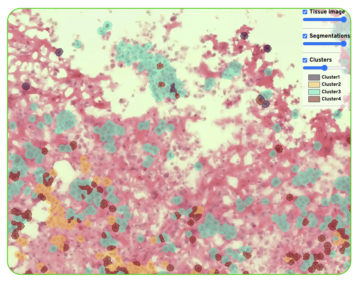
Our automated bioinformatics pipelines transition your project from raw imaging and sequencing metrics to profound biological insight. We employ industry-standard workflows tailored to multi-dimensional data—transforming complex count matrices and image alignments into clear, interactive biological landscapes using tools like Space Ranger, Xenium Ranger, Seurat, and Squidpy.
Deliverables Include
- Transcript overlays on H&E scans
- Cell segmentation
- UMAP of segmented cells
- Graph-based cluster tissue plots
- Spatial expression tissue mapping
- Neighborhood analysis
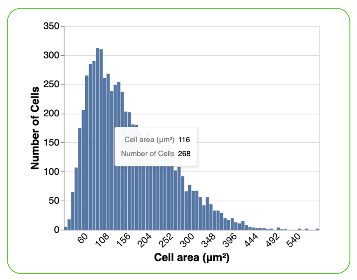
- Cell size distribution
- UMI count to image alignment

Cell Segmentations

Cell Size Distribution
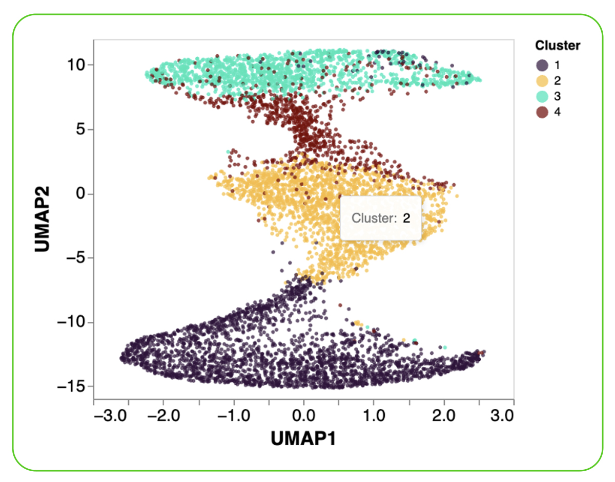
UMAP of Segmented Cells
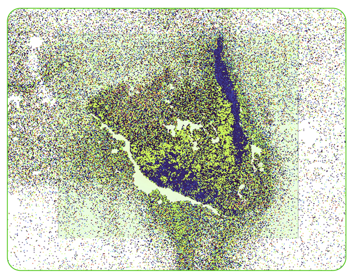
Tissue Plot with 8 µm Bins Colored by Graph-based Clustering
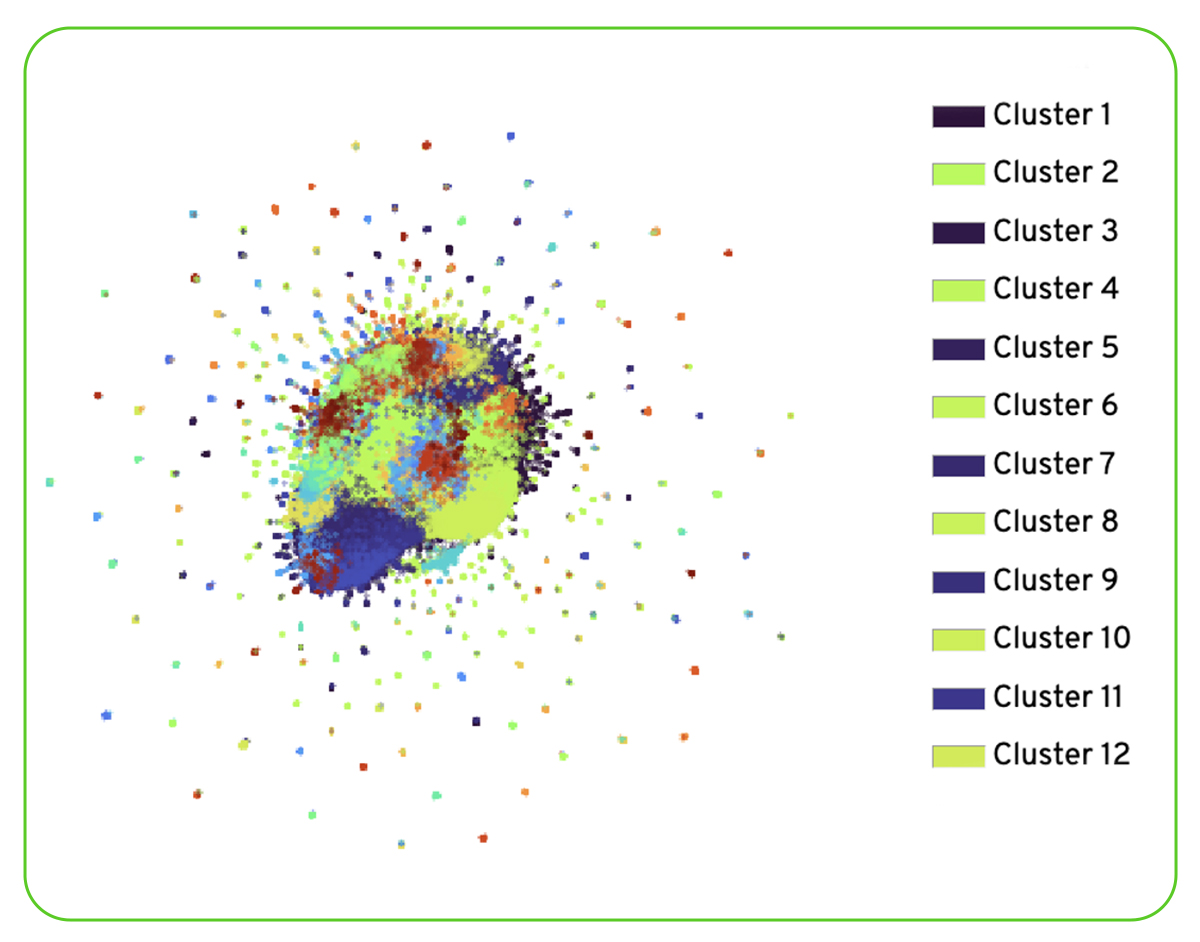
UMAP Projection of 8 µm Bins Colored by Graph-based Clustering
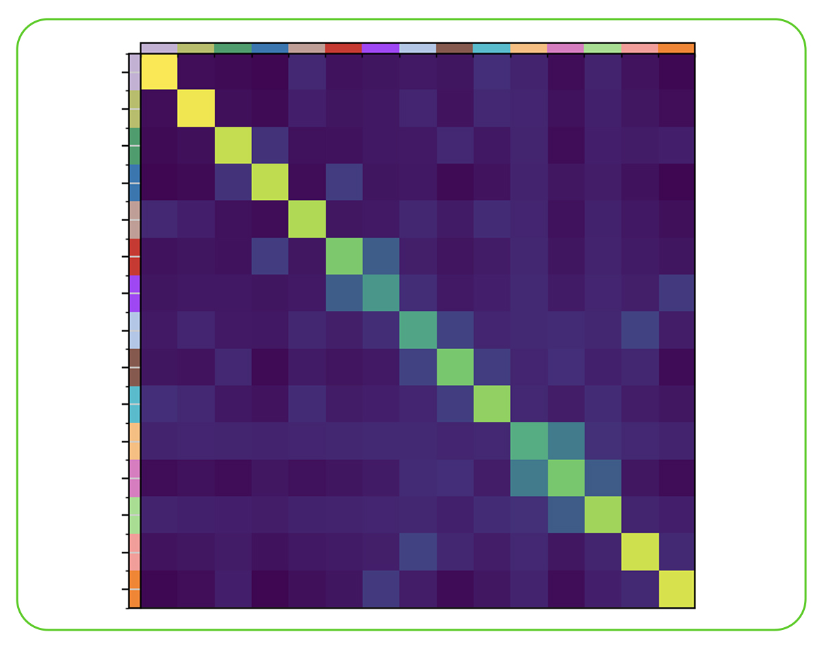
Neighborhood Analysis
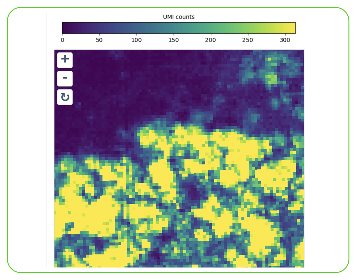
Total UMI Count to Image Alignment
Applications of Spatial Transcriptomics Sequencing
Our high-level spatial biology services support a broad range of multi-disciplinary applications, including:

Tumor Microenvironment (TME) Architecture
Map exact coordinates of immune cells, stromal barriers, and tumor margins to identify local patterns of immune evasion and infiltration.

Neurobiology & Brain Mapping
Resolve the intricate organization of cellular layers in central nervous system tissues and track regional gene expression across distinct brain structures.

Biomarker Validation & Translational Screens
Validate expression patterns of therapeutic drug targets and investigate structural changes across clinical tissue cohorts pre- and post-treatment.

Drug Mechanism of Action
Evaluating how novel therapeutic compounds affect cellular pathways across various tissues while maintaining physiological morphology context.

Resources & Insights
![]()
SERVICE OVERVIEW | TECH SHEET
Spatial Transcriptomics Services
Download the technical overview of our spatial workflows to see how we map high-resolution gene expression directly within tissue sections while preserving structural morphology.
![]()
Blog
Visium vs. Xenium vs. StrataMap: Choosing the Right Spatial Resolution
Learn how to balance whole-transcriptome discovery against subcellular targeted resolution to maximize research ROI based on sample dimensions and budget.
![]()
White Paper
Maximize Discoveries with Spatial Transcriptomics
A concise overview of spatial transcriptomics and its key applications in developmental biology, neuroscience, cancer research, and regenerative medicine.
Frequently Asked Questions
Planning your Spatial Project
How do I initiate a spatial transcriptomics project?
Click our Request a Quote link or reach out through our portal. Our specialists will schedule an introductory technical call to discuss tissue block processing, slide compatibility, and provide your project quote.
What is the main operational difference between Visium, Xenium, and StrataMap?
Visium and StrataMap are both unbiased, whole-transcriptome discovery platforms. Visium offers traditional spot-based next-generation spatial workflows. StrataMap leverages a massive 750mm² capture area with continuous 1µm surface features to achieve high-sensitivity, single-cell binned whole-transcriptome mapping. Xenium, by contrast, focuses on targeted in situ single-molecule panels for precise, subcellular cellular classification.
What sample types are compatible across your platforms?
Our services are validated for an extensive range of inputs, primarily focusing on Formalin-Fixed Paraffin-Embedded (FFPE) blocks and fresh-frozen (FF) tissues. Compatibility varies slightly by platform; our team will verify your specific tissue type during your initial consultation.
What bioinformatics options are provided for spatial datasets?
Our spatial analysis includes automated image alignment, transcript mapping, spatial cluster generation (Space Ranger/Xenium Ranger), tissue zone partitioning, cell segmentation adjustments, and interactive visualization files.
What tissue formats and preparation states are supported?
We support custom spatial workflows optimized for both Fresh-Frozen (FF) blocks embedded in OCT and Formalin-Fixed Paraffin-Embedded (FFPE) tissue blocks prepared using standard clinical histology methods.
Are there any specific tissue-quality metrics required before starting?
Yes. For fresh-frozen tissue, we recommend RIN >7. For FFPE blocks, we assess quality using a DV200 >30%, ideally targeting DV200 >50% for optimal assay performance.
What is the typical turnaround time (TAT)?
Typical turnaround times range from 4 to 6 weeks from sample receipt to final data delivery, depending on the scale of the project, sample quality, and chosen sequencing depth.
Can you handle sectioning, or do I need to send pre-cut slides?
We offer complete, end-to-end processing. You can send us your raw tissue blocks, and our certified in-house histology core will handle precision sectioning, slide placement, quality testing, and staining.
Explore Related Sequencing Solutions
High-resolution transcriptomics to resolve individual cellular heterogeneity.
Resolving complex structural variations and full-length RNA isoforms.
High-plex protein profiling for functional multi-omic integration.
Get started with your project
Ready to accelerate your spatial biology research? Contact our team for a technical consultation. We will guide you through sample requirements, technology options, slide architectures, and library prep aligned to your analysis goals.
- 10x Genomics Certified Service Provider
- In-house sectioning, H&E, and TMA construction
- Visium, Xenium & Illumina StrataMap
- Publication-ready spatial figures
Request a quote
