
Feature Linkage: The Power of Same-Cell Covariation
The 10x Genomics Multiome workflow removes this ambiguity. By adding cell-specific barcodes to both transposed genomic DNA fragments and nuclear mRNA inside the exact same droplet partition, we capture an unedited view of cell-by-cell covariation.
In downstream data processing, our pipelines move beyond simple coordinate matching to calculate explicit Feature Linkages. This algorithm measures the absolute covariation between targeted genes and proximal open chromatin peaks across thousands of individual cells.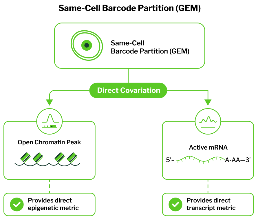
Because both measurements share an identical cell barcode, we can employ advanced regression frameworks—such as regularized Poisson regression tiles—to model the collective impact of all surrounding accessible areas simultaneously. This approach allows us to differentiate true regulatory elements from secondary, non-causal “tagging” peaks that merely open in tandem due to shared transcription factor binding.
What Our Pilot Data Reveals: Uncovering Hidden Regulatory Elements
Benchmarking evaluations across our internal Multiome control cohorts highlight exactly what researchers miss when running separate assays.
When analyzing standard peripheral blood mononuclear cells (PBMCs) and complex tumor biopsies, traditional unlinked analyses routinely fail to detect key distal regulatory nodes. The figure below highlights a representative genomic locus where a critical, cell-type-specific enhancer is masked by standard separate processing but cleanly resolved via single-cell Multiome profiling.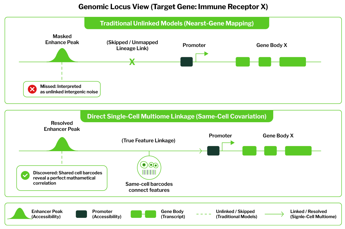
(Discovered: Shared cell barcodes reveal a perfect mathematical correlation)
By tracking precise cell-by-cell covariation, our pipelines mapped an open chromatin cluster located over 120 kilobases upstream directly to the promoter of a major immune receptor. Traditional, separate workflows had previously discarded this peak as unlinked intergenic noise because its opening sequence did not match the expression patterns of the immediately adjacent, non-target gene body.
Multiome analysis proved that this distal peak served as a key lineage enhancer, opening exclusively within a rare subset of activated T cells immediately prior to target gene transcription.
Transforming Biomarker Discovery and Drug Screening
Transitioning from inferred computational models to true multiomic resolution alters the scope of translational research:
- Fine-Mapping Disease Variants: Researchers can intersect patient-derived GWAS datasets directly with verified Feature Linkage maps. This immediately identifies whether a non-coding risk variant physically sits within an active, tissue-specific enhancer loop that controls a downstream disease driver.
- Characterizing Epigenetic Pioneer Drivers: Identify exact transcription factor motifs embedded within newly opened enhancers, defining the precise cascade of molecular switches that govern drug resistance, cellular differentiation, or malignant transformations.
- Accelerating Therapeutic Targeting: Instead of screening drugs based solely on broad transcriptomic shifts, investigators can monitor whether small-molecule inhibitors effectively close specific, remote enhancer regions, stopping disease-driving gene networks at their epigenetic root.
Accelerate Your Multiomic Insights with Signios Bio
Capturing these interconnected regulatory layers requires exceptional sample processing. Isolating single nuclei while preserving clean structural chromatin envelopes and fragile nuclear mRNA necessitates rapid, temperature-controlled laboratory execution.
At Signios Bio, our Foster City, CA specialists handle this entire technical sequence for you. From running automated tissue dissociation and specialized detergent washes to executing multi-library separations and deep sequencing on our Illumina NovaSeq platforms, we optimize every parameter to maximize your Transcription Start Site (TSS) enrichment scores.
You deliver the tissue or frozen specimens; we deliver the fully integrated, publication-ready multiomic matrices and peak linkage profiles your discovery pipeline demands.