
By Scientific Affairs Team, Signios
When your entire experiment relies on a few hundred laser-captured cells, a rare circulating tumor cell population, or a handful of precious fluorescence-activated cell sorting (FACS) isolates, the stakes at the laboratory bench are exceptionally high. With ultra-low input and single-cell RNA sequencing (RNA-Seq), traditional rules of sample preparation do not apply. A single misstep during collection won’t just lower your yield—it can erase your entire sample.
To ensure your trace biological samples yield robust, publication-ready libraries, you must actively protect your RNA from the moment of isolation. This guide breaks down the critical technical parameters required to safeguard low-biomass samples before they reach the sequencer.
1. The Low-Input Enemy: Traditional Column Extraction
For standard sequencing workflows, purifying RNA through a silica-membrane column is the gold standard. However, when working with inputs below 1,000 cells or less than 10 nanograms (10 ng) of total RNA, traditional column extraction is the enemy of your data.
Silica membranes exhibit a natural binding pocket saturation threshold. When trace amounts of nucleic acids pass through the column, a significant percentage remains irreversibly bound to the membrane matrix.
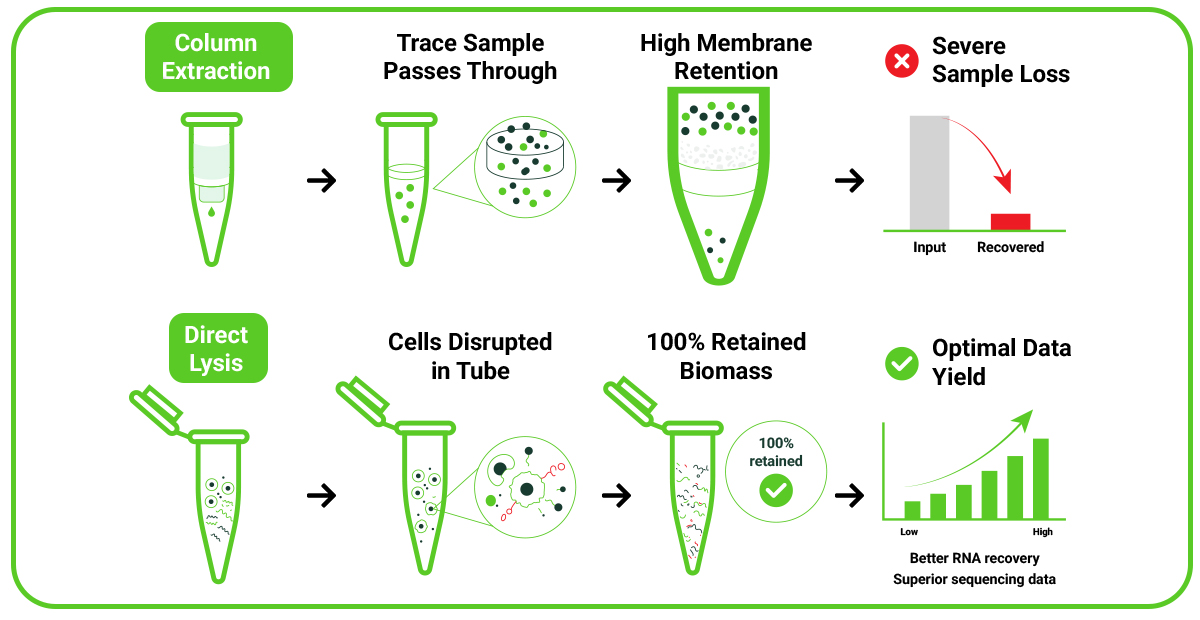
The Solution: Direct Cell Lysis
To circumvent this physical loss, our ultra-low input workflow utilizes direct cell lysis. Instead of isolating and eluting the RNA, cells are sorted or gathered directly into a highly concentrated, specialized lysis buffer.
This approach:
- Completely eliminates the wash-and-elute steps where precious material is lost.
- Instantly denatures endogenous RNases (enzymes that degrade RNA) upon cell contact.
- Preserves the entire cellular biomass in a single tube, ensuring that your starting material matches your analytical input.
2. Technical Parameters for Precise FACS Sorting
If your sample preparation involves FACS, how you handle the collection tube is just as critical as how you set your gating parameters. For ultra-low input workflows utilizing Takara SMART-Seq chemistry, adhere to these strict collection boundaries:
Keep Volumes Ultra-Low
Sort your cells into a microscopic volume—ideally 1 to 5 microliters ( μL ) of validated Signios lysis buffer.
Why volume matters: Downstream reverse transcription reactions occur in incredibly tight volume constraints (often under 20 μL). If your sorted cell volume is too large, it dilutes the reaction chemistry, severely reducing template-switching efficiency and hurting your final library complexity.
Calibrate the Stream
Ensure your sorting core is perfectly aligned. Off-target droplets that hit the dry upper walls of the PCR tube will evaporate, killing the cells and degrading the RNA before the lysis buffer can protect it. Drops must merge directly into the liquid buffer at the very bottom of the tube.